Highly realistic simulations for training could soon be widely used to rehearse complex cases prior to surgery, thanks to Simulated Inanimate Model for a Physical Learning Experience (SIMPLE), the brainchild of Ahmed Ghazi, M.D., M.Sc., an assistant professor in the Department of Urology at University of Rochester Medical Center, and Neurosurgery resident Jonathan Stone, M.D.
SIMPLE is a new new way to fabricate artificial organs and human anatomy that mimics the real thing, even up to the point of bleeding when cut. It entails converting images obtained from medical scans into computer generated designs and, through the assistance of 3D printing, fabricating lifelike organs that can be poked, prodded, and dissected.
The process begins with images obtained from MRI, CT, or ultrasound scans which then become computer-assisted designs (CAD). Rather than creating rigid plastic replicas of human anatomy, which was already being done in many other places, they converted the CADs of organs into molds, or negatives, which were built using a 3-D printer. The molds are then injected with a hydrogel which, after freezing, assumes a solid state. Since the water consistency of the hydrogel is identical to that found in our bodies giving the artificial organs the same feeling as the real thing.
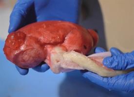
This artificial kidney is hydrogel injected into a 3D printed mold. Credit: University of Rochester
A flight simulator for surgery
Once the basic models of human anatomy were created, the pair began to tweak the designs in order to change the pathology. For example, they would alter the concentration of the hydrogel to add a denser tumor mass to a liver, or a blockage in a kidney, or plaque in an artery. Using the 3-D printer to create more rigid structures, the team can also create bone to simulate procedures involving the spine and skull.
Just being able to handle and examine a replica of a real organ can provide surgeons with a great deal of insight and information. They can observe where the blood vessels enter and leave the organ and, if it is a cancer model, the size and location of the tumor. They can even cut away at the organ to take a look at the interior. Now students, trainees, and surgeons can replicate the complete surgical experience, which required not only building the organs of interest, but the rest of the surrounding human anatomy so the entire surgical process of guiding instruments to the right location, moving other organs out of the way, clamping blood vessels, and resecting and removing tumors could be replicated.
To accomplish this feat, the team assembles entire segments of the body, complete with artificial muscle tissue, skin and fat, and, depending upon the area of interest, the liver, intestines, spleen, kidney, and other adjacent organs and structures. Artificial blood vessels are connected to bags of red dye that will “bleed” if cut. This was also done with other bodily fluids such as urine or bile.
Best consumer simulation ever next?
The assembled unit is then brought into the operating room where it is hooked up to a robotic surgical system, and the entire procedure simulated from the first insertion of instruments to completion.
The lifelike nature of the simulation has occasionally caused even trained professionals to do a double take.
“We have had times when we are doing these simulations in the OR when nurses or other physicians have looked in the window and thought we were doing the real thing, and have even gone so far as to scrub and put their masks on before coming in thinking there was a patient on the table,” said Ghazi.
While the simulations can be used to train on a generic model of anatomy, the ultimate vision is to harness this technology so that it can enable surgeons to rehearse complex cases before the patient is brought into the operating room. In these instances, the team can build organs using the actual patient scans, accurately replicating the unique conditions that will be found during the live operation.
Makes you want to try your own hand at simulated surgery, right? Surgery, like lots of things, often has a plan until things start. Which could make for a tense, thrilling experience for people willing to pay to try their hand at something without the life-altering consequences.
“Surgery is often like a Pandora’s Box,” said Ghazi. “You don’t know what is inside until you open it up. The fact that we could someday have surgeons practice procedures on these models before going to the operating room helps eliminate the unknown, increases safety, and improves the quality of care. Patients can, in turn, reassure themselves by asking their surgeons ‘how did the rehearsal go yesterday?’ That is going to be the future of surgery.”