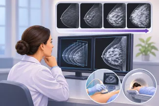
Have you been hearing about how your breast density may impact the risk of breast cancer? Conflicting information on this topic can be confusing and scary. So first, let’s get into what dense breast tissue actually means.
Our breasts are composed of three main components: fat, fibrous tissue, and glandular tissue. Fibroglandular tissue includes our milk ducts and lobules and shows up white on a mammogram. The fatty component appears dark gray or black on a mammogram.
Dense breast tissue is normal and common. Approximately half of all women above age 40 have dense breasts. The more fibroglandular tissue you have, the denser your breasts are and the more white the mammogram appears.
How do you know if you have dense breast tissue?
You cannot tell by sight or touch alone. Many women think that since their breasts feel lumpy during their self-exams, this must mean they have dense tissue, but that is not the case and is often why self-exams can lead to unwarranted concerns. Breast size or shape does not always correlate to density. A woman who wears a DD cup size may not have dense tissue and may have mostly fatty breasts. The best and only way to find out your breast density is via imaging such as a mammogram.
Age is the primary factor affecting breast density. Breasts tend to become less dense with age, so younger women tend to have denser breasts. Our breast density can vary over our lifetime due to several reasons, including weight fluctuations, pregnancy, hormone replacement, certain chronic medical conditions, or surgeries such as breast reductions. That said, breast density usually does not change drastically.
Why is breast density important?
Dense breast tissue is considered a separate risk factor for breast cancer. In a case-control study in 2007, women with 75% density (category D, extremely dense breast tissue) had a risk of developing breast cancer that was 4.7 times higher than those with less than 10% density ( category A).
The exact mechanism of why this is the case is not yet fully understood, but the reason is likely multifaceted, such as a combination of hormonal influence and having more tissue from which a cancer can grow.
A mammogram is the only way to determine breast density
The radiologist interpreting your mammogram will evaluate the breast density. Breast density falls into four categories, ranging from fatty or not dense (Category A) to extremely dense (Category D). The key concern is that mammogram sensitivity decreases as breast density increases. Overlying dense breast tissue can obscure or hide an underlying cancer because there is very little contrast between the tissue and the fat. Therefore, supplemental screening with ultrasound or MRI should be considered for those with category C or D breast density to refine, not replace, the mammogram’s findings.
Federal law requires doctors to notify patients if they see dense breast tissue on a mammogram. The radiologist will include this information under a special subsection detailing which breast density category is seen.
Mammograms are the gold standard in breast cancer screening
If mammograms are limited in dense breast tissue, should you skip them and get a different type of imaging instead? No. Mammograms are effective and considered the gold standard in breast cancer screening even in women with dense breast tissue. One major reason for this is that not all breast cancers appear as masses. Some, often those at the earliest stages, present as calcium deposits, calcifications that often appear only on mammograms and can be missed on ultrasound and MRI. The same is true for distortions, areas of “pulled” tissue that appear star-shaped on a mammogram and indicate an abnormal process within the breast tissue. This area, which stands out on a mammogram, may appear normal on an ultrasound.
Most mammograms today include 3D views, digital breast tomosynthesis, or 3D mammograms, which help the radiologist better interpret tissue because dense breast tissue can mask certain underlying cancers.
To maximize the benefits of mammography, compression is an important step in obtaining diagnostic-quality images. While compression can be the most uncomfortable part of a mammogram, it serves three major purposes. Compression helps flatten overlapping tissue, decreases image acquisition time and radiation dose, and holds the breast still so motion artifact from breathing or subtle movement does not prevent appropriate interpretation. Compression can help the radiologist detect subtle findings.
What other screening options exist?
Lifetime risk of developing breast cancer can be elevated in those with a family history of breast cancer, a genetic mutation such as BRCA mutations, or those who have received prior radiation to the chest. Risk stratification should be performed in all women between the ages of 25 and 30.
Reduction in deaths from breast cancer has only been studied in mammography. Therefore, additional screening in women with dense breast tissue is a point of contention among experts, and a standard has yet to be established.
According to the American Cancer Society, women with dense breast tissue can consider supplemental evaluation with ultrasound or MRI, as these imaging tests may help find some breast cancers that are not seen on mammograms. However, there are pitfalls with additional evaluation. For example, ultrasound evaluation depends on the skill and experience of the performing sonographer, MRI accessibility in rural areas may be limited, and additional imaging is not always covered by insurance and can incur additional costs. Supplemental evaluation can also lead to more findings and increased follow-up, testing, or biopsies that may not always be necessary. In women at greatest risk (20% or more), annual MRI is often recommended either as an independent screening tool in women less than age 40 or as a supplement to mammogram.
Being told you have dense breasts is neither a diagnosis nor a reason to panic. Breast density is common, normal, and simply one piece of the breast cancer risk puzzle. But it is an important piece because it creates a double challenge: increasing the likelihood of developing breast cancer while making it harder to detect. Mammograms remain the foundation of screening, even for women with dense breasts. The real value of knowing your breast density is not that it gives you another reason to worry, but that it gives you and your physician better information to make informed decisions.

Mansi Vaishnav, MD
Dr. Vaishnav is a board-certified breast radiologist, in a private practice, specializing in breast image interpretation and image-guided biopsies. She creates content to raise breast cancer awareness to reach more women and combat misinformation.