Inserting a camera into the human body, to look around for trouble, used to be the thing of science fiction. But as well all know full well, today millions of Americans visit doctor's offices annually to have a colonoscopy performed. It's become routine, and the cancer-spotting procedure saves lives.
Now instead of envisioning a camera that fits inside a colon, imagine one that can slide and peer through a blood vessel.
While you're imagining that, researchers in Michigan are currently experimenting with this new technology that may someday be able to predict a stroke or heart attack before it occurs.
A team of doctors at Michigan Medicine, the medical school at the University of Michigan in Ann Arbor, are using an incredibly small camera – it's called a scanning fiber endoscope, or SFE – to improve the view inside blood vessels and the carotid artery. The idea, when the technology is perfected, is to someday give doctors and surgeons a much clearer picture of blockages or potential obstructions that subsequently produce a cardiovacular event.
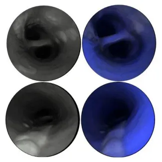
"The camera actually goes inside the vessels. We can see with very high resolution the surface of the vessels and any lesions, such as a ruptured plaque, that could cause a stroke," according to Dr. Luis Savastano, the study's first author. "This technology could possibly find the 'smoking gun' lesion in patients with strokes of unknown cause, and may even be able to show which silent, but at-risk, plaques may cause a cardiovascular event in the future."
According to Michigan Medicine, in a statement released today, "All research is in the pre-clinical phase."
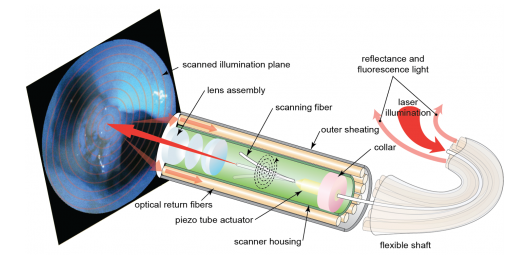 The technology, when you think about it, is quite amazing. Once the micro-camera is inserted, laser beams illuminate the area and high resolution images are gathered, then reassembled, and then returned along the outside of the device – in real time – for doctors to view. (Image courtesy of Michigan Medicine)
"Here, we demonstrate the use of a multimodal endoscope technology to collect laser-induced reflectance and fluorescence simultaneously, and perform structural and biological evaluation of atherosclerosis," states the study, published online today in the journal Nature Biomedical Engineering.
"This system consists of an ultrathin, highly flexible catheter that scans blue, green and red laser beams ... in a spiral pattern on the tissue surface, and that collects reflectance and fluorescence," according to the paper. By combining those two pieces, "we were able to co-generate endoscopic videos ... of healthy and diseased arteries."
The SFE's inventor is Dr. Eric Seibel, a mechanical engineering research professor at the University of Washington. A co-author of the paper, he originally designed the endoscope to improve early detection of cancer cells that could not be seen using available technology.
According to Dr. Seibel, who holds a Ph.D. – and, as the Michigan team disclosed, shares in the royalties generated from the device with U of W – adds that the device "can also assist neurosurgeons with therapeutic interventions by guiding stent placement, releasing drugs and biomaterials and helping with surgeries."
Given this wide-ranging potential, we look forward to the day when taking a peek inside a blood vessel becomes as routine as saying "ahhhhh" during one's annual physical.