Pregnancy ordinarily lasts about 40 weeks, but roughly one in ten babies worldwide is born prematurely (before 37 weeks of gestation). The consequences can extend well beyond the delivery room. Premature infants are at increased risk of respiratory disease, neurological injury, impaired vision and hearing, and developmental disabilities that may persist throughout their lives. The earlier the delivery, the greater those risks.
My wife and I are well versed in premature labor, spending multiple occasions during her first pregnancy in the Emergency Department, receiving fluids and medications, and increasingly her being asked to spend more time in bed. Through a combination of medication and luck, our first-born was ultimately born at term. However, he couldn’t pass up the opportunity to add one last “complication” to the pregnancy: a Cesarean section.
When Labor Begins Too Soon
A 2021 review of treatments for preterm birth estimated that approximately 15 million infants were born prematurely annually, with more than a million subsequently dying from complications within their first five years. Survival after very premature birth is dramatically better where neonatal intensive care is readily available, as is the case in the US.
The initiation of labor at term, like pregnancy itself, is a well-coordinated dance of many signals, meaning preterm birth isn't a single pathway but a common endpoint when the choreography is “off the beat.”
Those pathways involve a coordinated transformation of the uterus. Inflammatory signals increase, progesterone's ability to maintain uterine relaxation diminishes, and uterine smooth muscle cells become increasingly capable of contracting together. Prostaglandins help stimulate contractions and soften the cervix, while changes in the fetal membranes and cervix prepare the way for delivery. And this doesn't account for additional signaling from the almost-newborn.
There is no single treatment that will end this problem, and the medications so helpful for my wife cannot always reverse a process already underway. Most therapies used to delay premature labor buy relatively little time, often 48 hours or less. While invaluable, these therapies allow physicians to administer corticosteroids that accelerate fetal lung maturation or transfer the mother to a hospital equipped to care for a premature infant. They do not restore a pregnancy to its normal course.
This is why understanding the earliest cellular events that initiate labor matters. Rather than asking only how to suppress contractions, researchers are asking what causes uterine muscle cells to abandon their relatively relaxed state in the first place.
The Immune System's Unexpected Delivery: Immune Cells Supply the Power
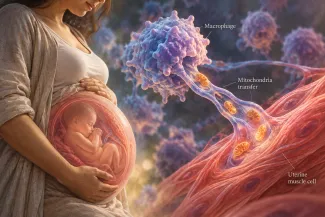
Among the many factors influencing the onset of labor is the mother’s immune system, which produces those inflammatory signals. A new study reveals a greater role than previously known. Monocytes are attracted to markers of injury and inflammation, where they can become macrophages, cells that engulf and remove pathogens, dead cells, and debris. Monocytes increasingly infiltrate the uterus during labor. Through a series of experiments, researchers extended our understanding of what happens next.
- Macrophages can adopt different functional states; the M1 form is pro-inflammatory, and the M2 form is anti-inflammatory and tissue-repairing. Near the onset of labor, macrophages shift toward the M1 phenotype, which establishes temporary physical connections, called tunneling nanotubes, and delivers intracellular cargo, including mitochondria, the body’s powerhouses, to uterine muscle cells. These muscle cells show increased ATP availability, the body’s fuel.
- The transfer is unidirectional: macrophages donate, and uterine muscle cells receive. A protein associated with tunneling nanotube formation was more abundant when macrophages were co-cultured with uterine muscle cells from women in labor, suggesting that these muscle cells may actively promote these connections, perhaps through their own chemical signals.
- Progesterone normally keeps the uterus quiescent, and its withdrawal is part of the initiation of labor. Although overall progesterone levels are maintained in human females, the researchers found that the same M1 cells showed changes consistent with both local progesterone withdrawal and activation of the uterine muscle. Mitochondrial transfer contributed to this process but did not account for all of it; other macrophage-derived signals appear to be involved. Macrophages do not merely contact uterine muscle cells; they help alter the molecular machinery that regulates their contractility.
- In a series of experiments on pregnant mice, the researchers showed that labeled macrophages and mitochondrial transfer increased before labor began, supporting the possibility that this contributes to preparing uterine muscle for contractions.
The emerging picture is that M1 macrophages may help prepare the uterus for labor in two complementary ways: by reducing progesterone's inhibitory influence on uterine contractions and by increasing the energy available to sustain them. While a compelling proposed mechanism, it is not a demonstrated cause of human labor or, more importantly, an established means of preventing preterm birth.
Treating Preterm Labor: Buying Time Rather Than Stopping the Clock
The new research offers a possible explanation for how inflammatory immune cells help prepare the uterus for labor. It may also help explain why pharmacologic treatment has been so frustrating. Most of our therapeutic armamentarium, the tocolytics, suppress contractions without necessarily reversing the biological changes that initiate them. These medications can:
- Relax the muscle using calcium channel blockers (an off-label use) that restrict calcium entry into muscle cells, an essential step in contraction. Alternatively, beta-adrenergic drugs directly relax smooth muscle cells. However, while they suppress contractions, they also increase heart rate, cause arrhythmias, and alter maternal blood glucose levels, limiting their use due to their adverse effect profile.
- Block the signals that promote contraction. In the US, indomethacin can be used because it inhibits prostaglandin production, which in turn stimulates uterine contraction and cervical softening. Unfortunately, its use, especially in early pregnancy, can have complications for the developing child, including impaired kidney function and constriction of the ductus arteriosus, an essential fetal blood vessel.
Our experience with progesterone, especially in light of this new study, helps explain the difficulty of defining treatment when one is still trying to understand a very basic physiologic process.
Throughout pregnancy, progesterone helps maintain uterine relaxation. Adding progesterone would seem like a logical way to prevent premature labor. Yet clinical trials have produced inconsistent results.
As the new mitochondrial-transfer study helps illustrate, labor in humans does not require circulating progesterone to disappear. Instead, uterine muscle cells can reduce progesterone's effectiveness locally, both by metabolizing the hormone and changing how its receptors respond. Adding more progesterone may be insufficient if the machinery that maintains its effects has already changed.
Newer experimental therapies that alter inflammatory signals aim to intervene upstream, before the uterus becomes fully activated. But suppressing inflammation involves a biological trade-off: the inflammatory response that contributes to premature labor may also protect the mother and fetus from infection.
For decades, preventing premature labor has largely meant trying to silence a uterus already preparing to contract. The new research suggests that the preparation itself may be a therapeutic target. By identifying how immune cells communicate with uterine muscle, alter progesterone signaling, and potentially supplement the energy needed for contractions, researchers have moved another step upstream in the chain of events leading to birth. Whether interrupting that communication can prevent premature delivery remains unknown. But it offers something our current pharmacology has largely lacked: another opportunity to intervene before labor is fully underway.
Source: Macrophage to Myocyte Mitochondrial Transfer in the Myometrium: A Novel Mechanism for the Initiation of Labor BioRxiv DOI: 10.64898/2026.08.17.745259
Landscape of Preterm Birth Therapeutics and a Path Forward Journal of Clinical Medicine DOI: 10.3390/ jcm10132912

Chuck Dinerstein, MD, MBA
Director of Medicine
Charles Dinerstein, MD, MBA, is the Medical Director of the American Council on Science and Health. A vascular surgeon with more than 25 years of clinical experience, he brings a physician's understanding of medical decision-making, an appreciation for scientific uncertainty, and a broader perspective on healthcare economics and policy to his writing.