For the last 100 years, slicing a single cell into two equal parts has proven to be a process that's tedious, time-consuming and one that required to be done by hand.
But a young, observant scientist and her fellow researchers at Stanford University have just come up with a method that's 200 times faster, with similar survival rates, and one, according to the school that "could eventually help scientists study and treat a variety of human diseases related to cell regeneration, such as cancer."
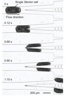
The researchers, led by Sindy Tang, an assistant professor of mechanical engineering, have created a "microfluidic guillotine" that channels single cells down a tight corridor before they are bisected by a pointed blade, creating two evenly-sliced cells (see adjacent graphic, courtesy of Lucas R. Blauch et al, Stanford University Dept. of Mechanical Engineering). Since the early 20th century this type of cell slicing has been done by hand, under a microscope.
 "Cutting a single cell by hand takes about 3 minutes if you're good at it, and even if you're good at it, you can't always cut the cell equally in half. This method has not changed for over 100 years," according to Lucas Blauch, a graduate student on Tang's team. "We knew that our lab's expertise in microfluidics would allow us to create a device to do that much faster."
Ms. Tang was prompted toward this innovation while learning about a single-celled, free-living freshwater organism known as the Stentor coeruleus, which, if it were to be cut in two, can heal itself into a pair of healthy cells. The Stentor was used in this work, the authors wrote in their paper, published online in the Proceedings of the National Academy of Sciences, "due to its robust repair capacity and the ability to perform gene knockdown in a high-throughput manner."
"It is one of the Holy Grails of engineering to make self-healing materials and machines," states Ms. Tang, in yesterday's news release from Stanford. "A single cell is analogous to a spacecraft – both have to figure out how to repair damage without anyone's help from the outside."
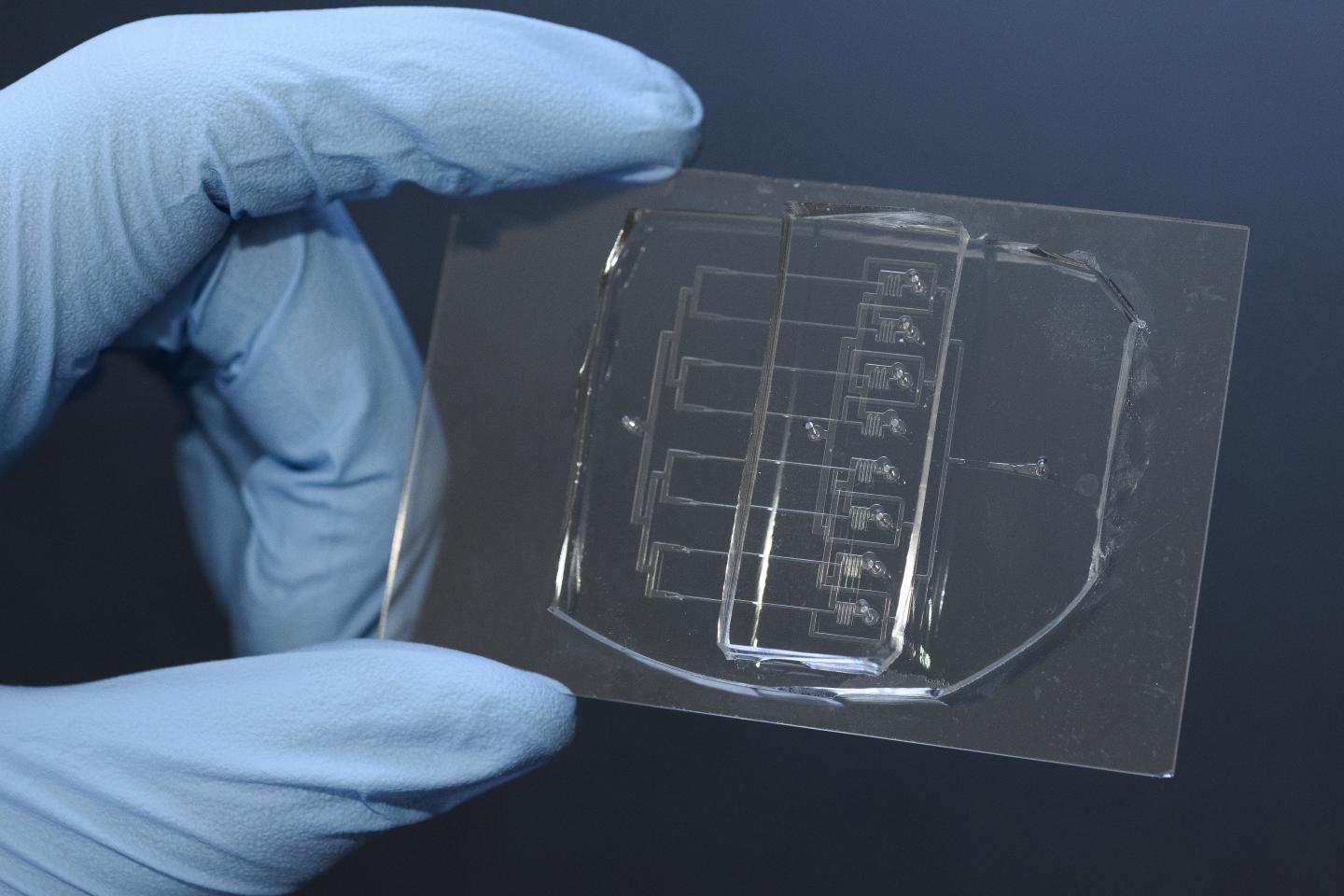
Once the research team devised the method for bisection, it multiplied its productivity by creating a tool with eight parallel corridors, one that can be held between one's thumb and forefinger (see photo, credit: L.A. Cicero/Stanford News Service.)
 Studying how cells heal themselves potentially has a wide application in medical research, and the new splitting technique holds additional promise.
"Our microfluidic platform presents an important step toward such a standardized assay and is expected to lay the foundation for understanding how single cells repair themselves," the authors wrote. "Furthermore, it should be possible to adapt our microguillotine design to other cell types."