
Identifying skin cancer
There are a variety of cancers that involve our skin. Basal Cell cancers (BCC), although rarely fatal, are the most common worldwide cancers. Squamous cell cancers (SCC) are more aggressive, with devastating health consequences via local growth. Malignant melanoma, a tumor of the color-bearing skin cells, is the leading cause of women's death in the 25-45 age range.
While a careful examination of the skin has been the mainstay of diagnosis, diagnostic imaging has improved the physician's ability to discern benign from malignant. That matters because, ultimately, the diagnosis rests with a biopsy, the surgical removal of the tissue, and we do not want to miss an atypical tumor, nor do we want to remove every harmless skin lesion. Current ultrasound systems have 98% accuracy in discriminating between these tumors.
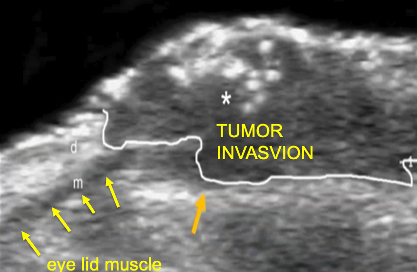 Once again, ultrasonography advances have improved diagnostic accuracy and often flag a biopsy on a visually innocent appearing lesion such as atypical melanoma or pigmented basal cell cancer. Specific echoes generated by nests of tumor cells are strong indicators of the cellular "aggression" that accompanies cancers. These 3-D images guide biopsy to the most virulent, "suspicious" area of the dermal tumor and also aid in documenting the extent of both the local lesion and spread to accompanying lymph nodes.
Treatments
There is a range of techniques to treat skin cancers. Topical therapies include chemotherapeutic agents like 5-fluorouracil cream and imiquimod for low risk, superficial lesions. Deeper lesions require various excisional methods. Perhaps the best known is Mohs micrographic surgery, where small slices of tissue are taken and immediately looked at under the microscope to determine whether cancer cells remain present or the "margin" is free of these cells. Advances in laser/optical devices allow near-microscopic tissue analysis of the cells by rapid, non-invasive testing and may well reduce the time required for this exacting surgical excision. Lasers have also been utilized in the destruction of these cancers. In these instances, high-resolution ultrasound is used to determine pre-treatment, the depth of the laser's destructive beam. Malignant melanoma is a far more aggressive, systemic disease. Immunotherapies have proven to be highly effective in improving survival. Some melanomas that are less than 1 mm in depth have greater survival and less invasive therapeutic course.
Lastly, superficial radiation therapy or SRT was a technique widely used in the early 1900s to treat skin cancers other than melanoma. While it was supplanted by Mohs surgery, it is mounting a bit of a resurgence as image-guided radiation techniques provide similar outcomes with less cosmetic alterations.
Sources: Neoplastic Diseases James Ewing
Image Guided Dermatology Treatment